|
One score will be assigned per root surface. The
facial, mesial, distal and lingual root surfaces of each tooth
should be classified as follows:
|
Code E
If the root surface cannot be
visualized directly as a result of gingival
recession or by gentle air-drying, then it is
excluded. Surfaces covered entirely by calculus
can be excluded or, preferably, the calculus can
be removed prior to determining the status of
the surface. Removal of calculus is recommended
for clinical trials and longitudinal studies. |
|
|
|
|
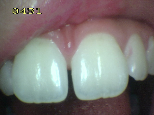
Code 0
The root surface does not exhibit any
unusual discoloration that distinguishes it from the
surrounding or adjacent root areas nor does it exhibit a
surface defect either at the cemento-enamel junction or
wholly on the root surface. The root surface has a
natural anatomical contour, OR The root surface may
exhibit a definite loss of surface continuity or
anatomical contour that is not consistent with the
dental caries process.
This loss of
surface integrity usually is associated with dietary
influences or habits such as abrasion or erosion. These
conditions usually occur on the facial surface. These
areas typically are smooth, shiny and hard. Abrasion is
characterized by a clearly defined outline with a sharp
border, whereas erosion has a more diffuse border.
Neither condition shows discoloration.
| |
|
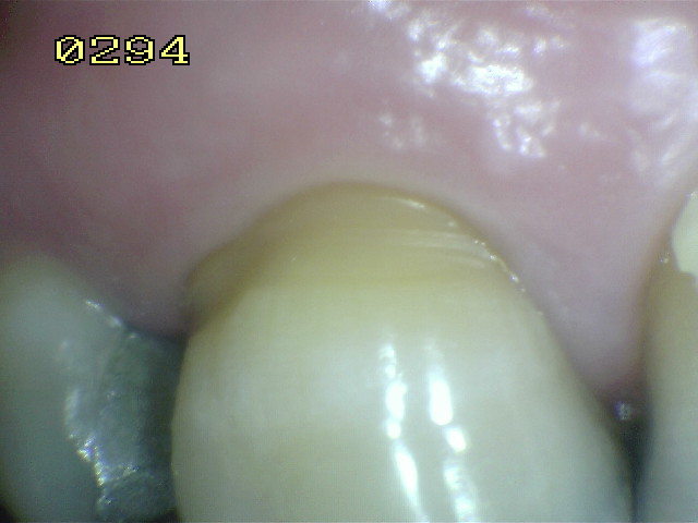
Code 1
There is a clearly demarcated area on
the root surface or at the cemento-enamel junction (cej)
that is discoloured (light/dark brown, black) but there
is no cavitation (loss of anatomical contour < 0.5 mm)
present. |
|
 |
|
Probe |
|
|
|
|
|
|
|
|
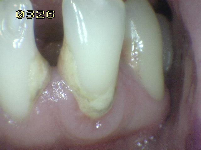
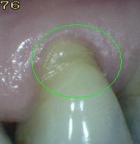
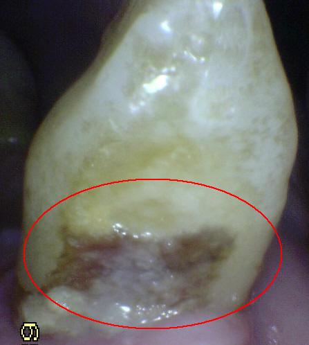
Code 2
There is a clearly demarcated
area on the root surface or at the cemento-enamel
junction (cej) that is discoloured (light/dark
brown, black) and there is cavitation (loss of
anatomical contour = 0.5 mm) present.
|
|
|
|
|
|
Probe |
|
|
|
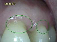
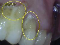
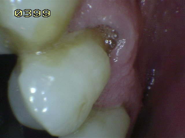
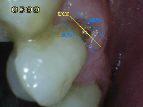
Special
considerations in the coding of root caries:
-
When the surface
of the crown and root are affected by caries they
must be identified independently. In case of doubt
because the caries lesion is in the cement-enamel
junction (UCE), it must be analyzed which surface is
more affected or which extends at least 1 mm or
beyond the limit of the cement enamel junction (UCE),
In both cervico-incisal and cervical apical
directions, it should be considered which is the
most extensive applying the 50% rule, if there is
equality the examiner must decide if the lesion is
coded as root or crown, or in its defect can apply
both . See right image.
|
 |
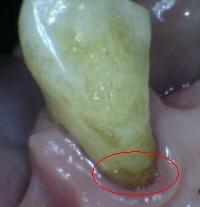
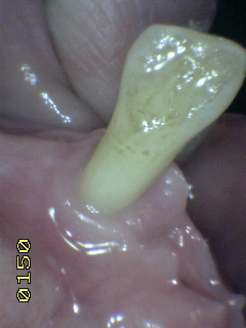
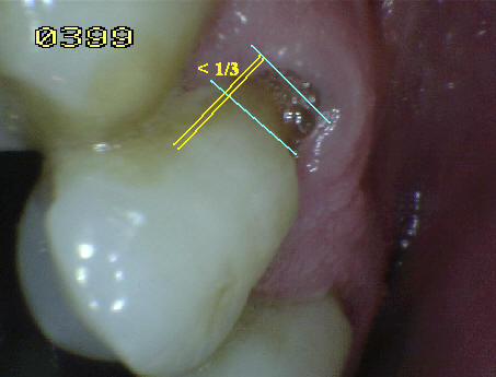
- When a carious lesion on a
root surface extends beyond the angle of the root
line but involves at least 1/3 of the distance
across the adjacent surface, that adjacent surface
must also qualify as caries. If it is smaller (<1/3)
it will be coded as sound. See right image.
- A root surface adjacent to a
rim of the crown that is free of decay should be
noted as sound.
- If more than one lesion is
present on the surface of the same root, the most
serious injury will be noted.
- All surfaces of the root
remains should be coded as "06".
- The non-vital teeth have the
same score as the vital teeth.
|
 |
The following diagram (Figure
1) will serve as a useful prompt for examiners in
deciding on appropriate coding of root caries:
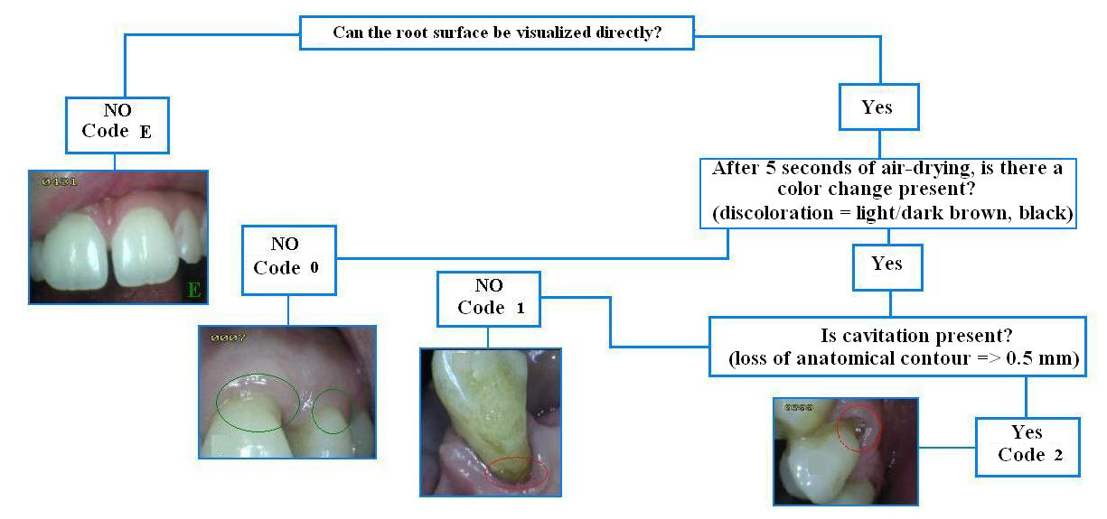
Figure 1: Decision tree for primary caries on thr root surface.
Caries associated with root restorations
When a root surface is filled and there is caries
adjacent to the restoration, the surface is scored as caries. The
criteria for caries associated with restorations on the roots of
teeth are the same as those for caries on non-restored root surfaces.
The following diagram (Figure 2) will assist the examiner in
deciding on the appropriate coding of caries adjacent to
restorations on root surfaces:
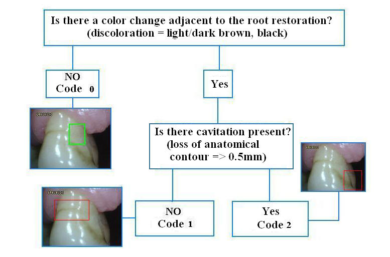
Figura 2: Decision treefor caries associated with root
restorations
Root caries activity:
The characteristics of the base of the discolored
area on the root surface can be used to determine whether or not the
root caries lesion is active or not. These characteristics include
texture (smooth, rough), appearance (shiny or glossy, matte or non-glossy)
and perception on gentle probing (soft, leathery, hard). Active root
caries lesions are usually located within 2mm. of the crest of the
gingival margin
The following diagram (Figure 3) will be helpful in
making a determination regarding the activity of root caries:
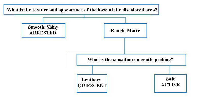
Figure 3: Decision tree for root caries activity |