|
Sound
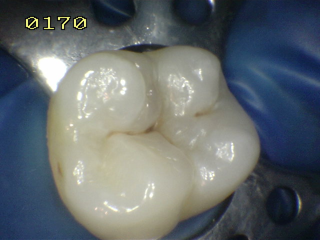
tooth surface: Code 0
There should be no evidence of caries (either no
or questionable change in enamel translucency after prolonged
air drying (suggested drying time 5 seconds)). Surfaces with
developmental efects such as enamel hypoplasias;
fluorosis; tooth wear (attrition,
abrasion and erosion),
and extrinsic or intrinsic stains will be recorded as sound.
The examiner should also score as sound a surface with multiple
stained fissures if such a condition is seen in other pits and
fissures, a condition which is consistent with non-carious
habits (e.g. frequent tea drinking) |
|
|
|
|
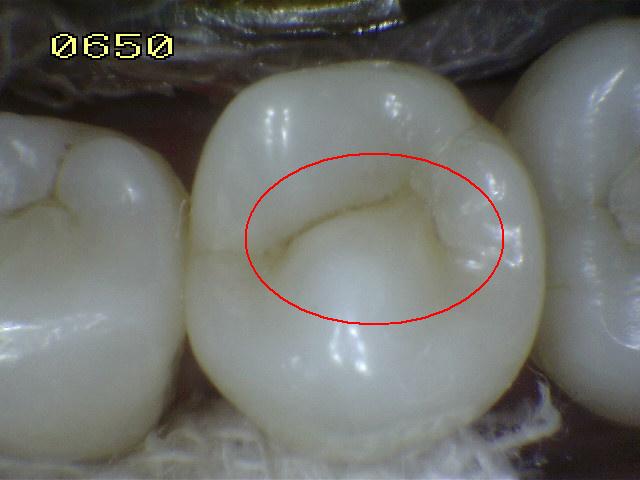
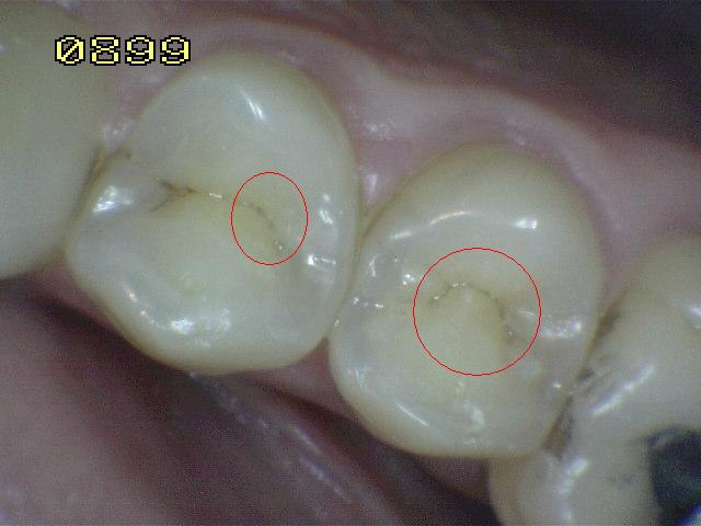
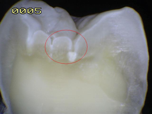
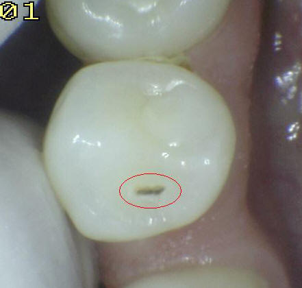
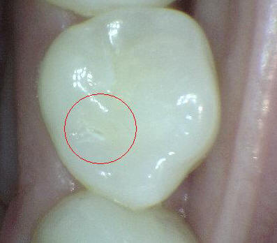
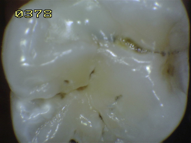
First visual change in enamel: Code 1
Code 1 is assigned for the following pits and fissures:
When seen wet there is no evidence of any change in color attributable
to carious activity, but after prolonged air drying (approximately 5
seconds is suggested to adequately dehydrate a carious lesion in enamel)
a carious opacity or discoloration (white or brown lesion) is visible
that is not consistent with the clinical appearance of sound enamel OR
When there is a change of color due to caries which is not consistent
with the clinical appearance of sound enamel and is limited to the
confines of the pit and fissure area (whether seen wet or dry). The
appearance of these carious areas is not consistent with that of stained
pits and fissures as defined in code 0.
|
|
|
|
|
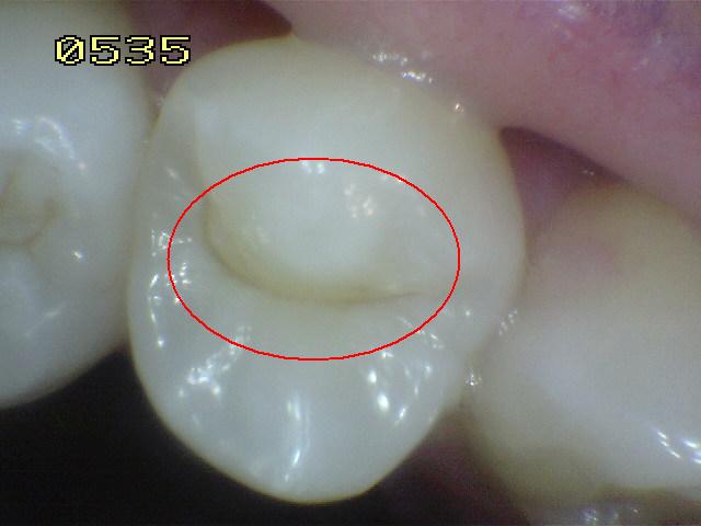
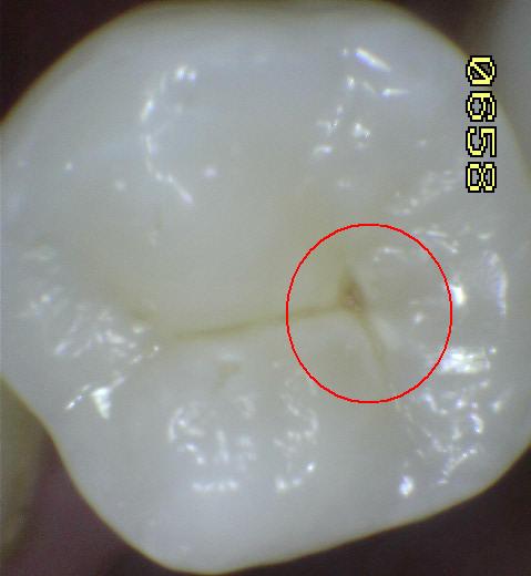
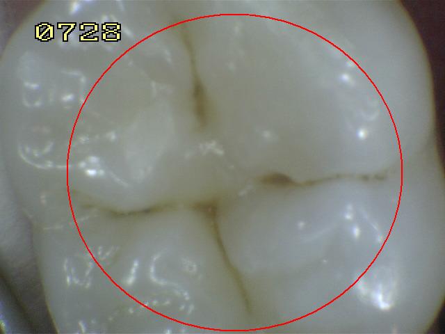
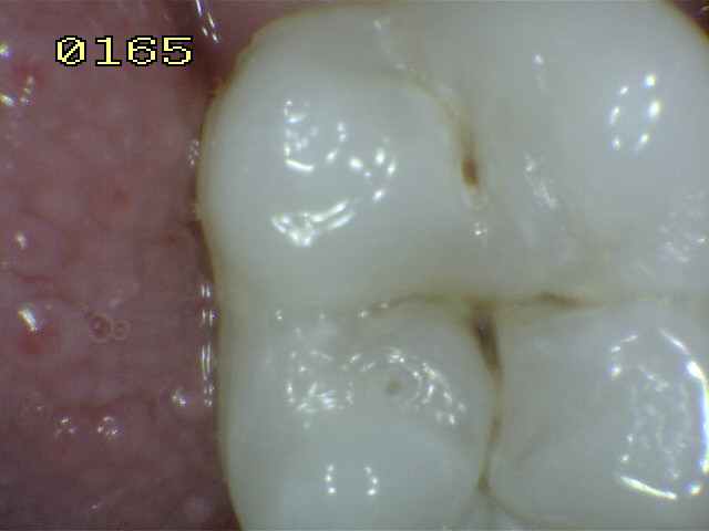
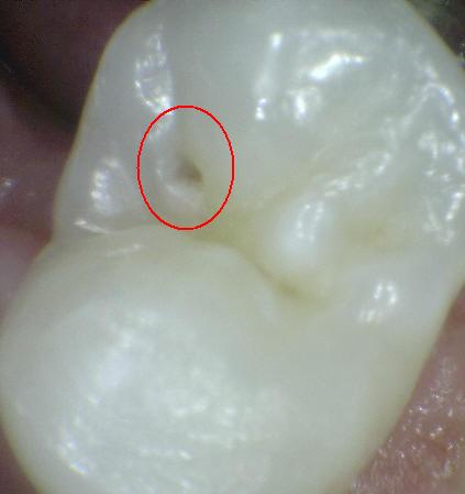
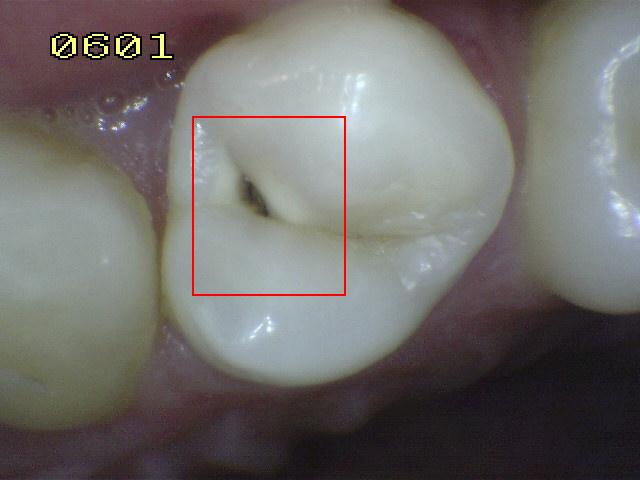
Distinct visual change in enamel: Code 2
The tooth must be viewed wet. When wet
there is a (a) carious opacity (white spot lesion) and/or
(b) brown carious discoloration which is wider than the
natural fissure/fossa that is not consistent with the
clinical appearance of sound enamel (Note: the lesion
must still be visible when dry).
|
|
|
|
|
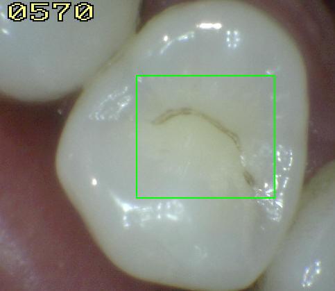
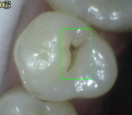
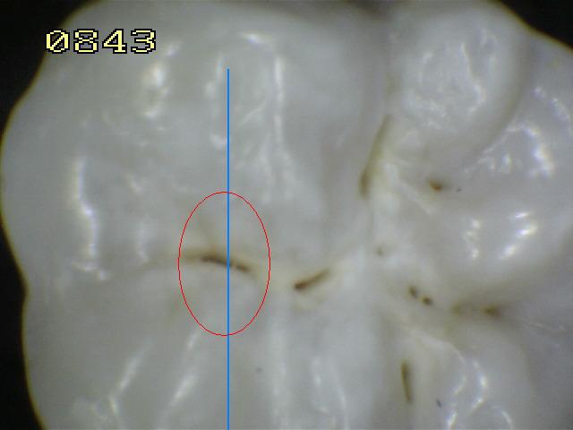
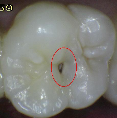
Localized enamel
breakdown due to caries with no visible dentin or underlying shadow:
Code 3
The tooth viewed wet may have a clear carious opacity
(white spot lesion) and/or brown carious discoloration which is wider
than the natural fissure/fossa that is not consistent with the clinical
appearance of sound enamel. Once dried for approximately 5 seconds
there is carious loss of tooth structure at the entrance to, or within,
the pit or fissure/fossa. This will be seen visually as evidence of
demineralization (opaque (white), brown or dark brown walls) at the
entrance to or within the fissure or pit, and although the pit or
fissure may appear substantially and unnaturally wider than normal, the
dentin is NOT visible in the walls or base of the cavity/discontinuity.
If in doubt, or to confirm the visual assessment, the
WHO/CPI/PSR probe can be used gently across
a tooth surface to confirm the presence of a cavity
apparently confined to the enamel.
This is achieved by sliding the ball end along the
suspect pit or fissure and a limited discontinuity is detected if the
ball drops into the surface of the enamel cavity/discontinuity.
|
|
 |
|
Probe |
|
|
|
 |
 |
|
IN VITRO |
|
|
|
|
|
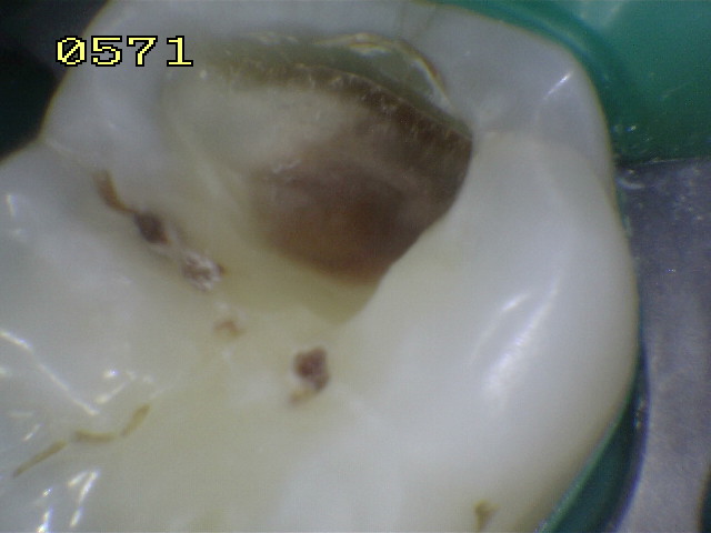
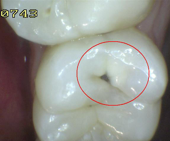
Underlying dark shadow from dentin with or
without localized enamel breakdown: Code 4
This lesion appears as a shadow
of discolored dentin visible through an
apparently intact enamel surface which may or
may not show signs of localized breakdown (loss
of continuity of the surface that is not showing
the dentin). The shadow appearance is often seen
more easily when the tooth is wet. The darkened
area is an intrinsic shadow which may appear as
grey, blue or brown in color. The shadow must
clearly represent caries that started on the
tooth surface being evaluated. If in the opinion
of the examiner, the carious lesion started on
an adjacent surface and there no evidence of any
caries on the surface being scored then the
surface should be coded “0”.
Code 3 and 4, histologically may
vary in depth with one being deeper than the
other and vice versa. This will depend on the
population and properties of the enamel. For
example more translucent and thinner enamel in
primary teeth may allow the undermining
discoloration of the dentin to be seen before
localized breakdown of enamel. However, in most
cases code 4 is likely to be deeper into dentin
than code 3.
|
Probe |
|
 |
 |
|
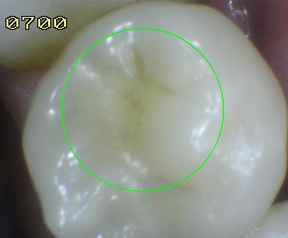
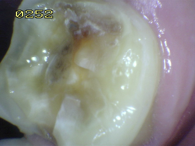
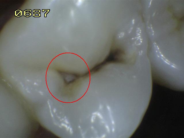
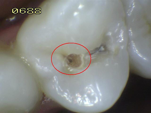
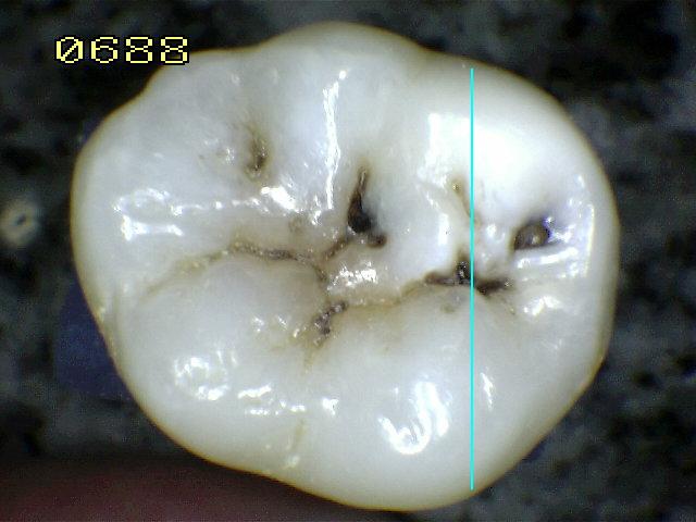
In the
image 0687, a code 4 and in the image 0688, a
caries in the dentine is observed after the
opening of the well with a crack. |
|
 |
 |
|
 |
 |
 |
|
 |
 |
 |
|
|
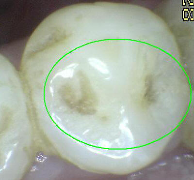
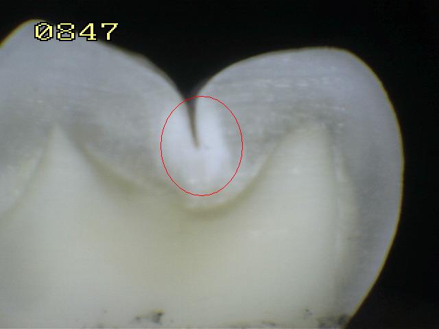
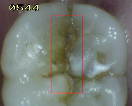
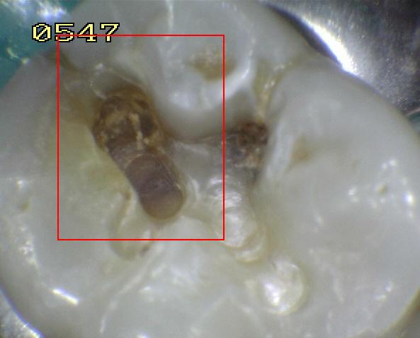
In the
image 0544, a code 4 and in the image 0547, a
decay of the dentine is observed after the
opening of the well with a crack. |
|
|
|
 |
 |
|
|
|
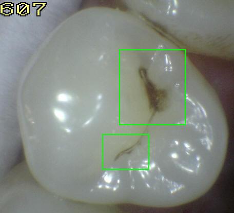
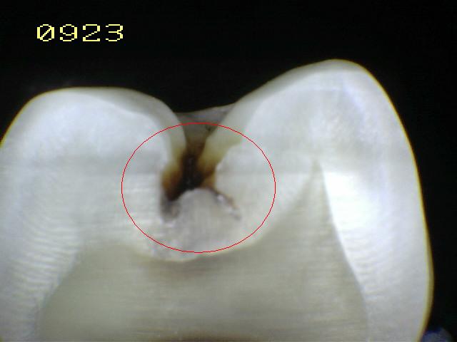
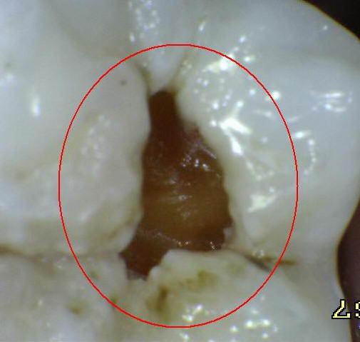
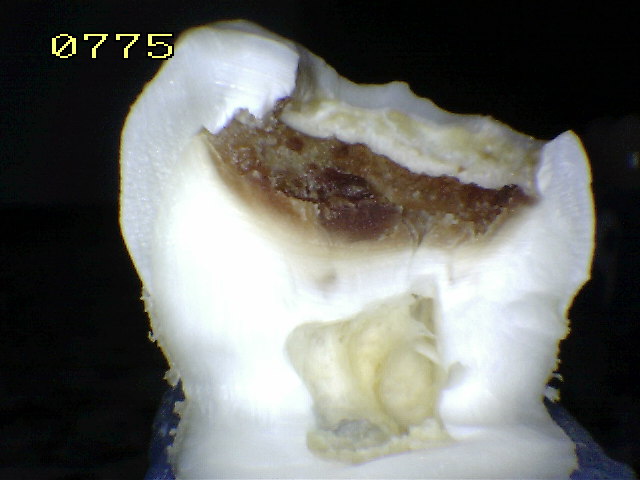
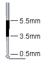
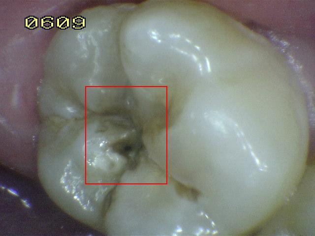
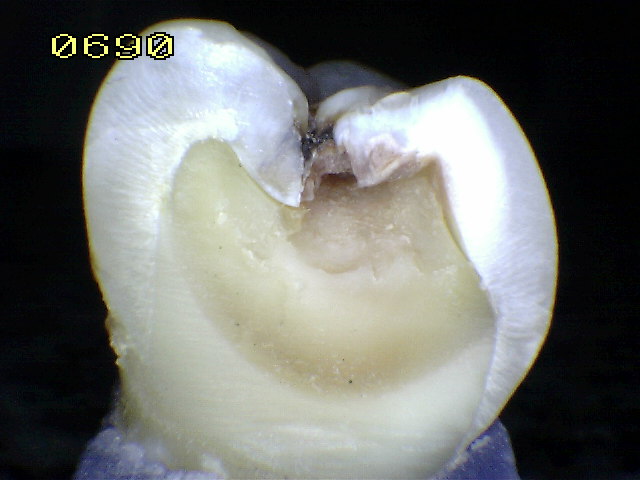
Distinct cavity with visible dentin: Code 5
Cavitation in opaque or
discolored enamel exposing the dentin beneath.
The tooth viewed wet may have darkening of the
dentin visible through the enamel. Once dried
for 5 seconds there is visual evidence of loss
of tooth structure at the entrance to or within
the pit or fissure – frank cavitation. There is
visual evidence of demineralization (opaque (white),
brown or dark brown walls) at the entrance to or
within the pit or fissure and in the examiner
judgment dentin is exposed.
The WHO/CPI/PSR probe can be
used to confirm the presence of a cavity
apparently in dentin. This is achieved by
sliding the ball end along the suspect pit or
fissure and a dentin cavity is detected if the
ball enters the opening of the cavity and in the
opinion of the examiner the base is in dentin.
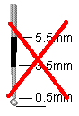
(In pits or fissures the thickness of the enamel
is between 0.5 and 1.0 mm. Note the deep pulpal
dentin should not be probed) |
|
|
|
Probe |
|
|
|
IN VITRO |
|
 |
 |
|
|
|
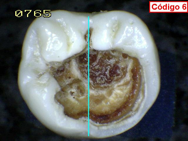
Extensive distinct cavity with visible dentin:
Code 6
Obvious loss of tooth structure,
the cavity is both deep and wide and dentin is
clearly visible on the walls and at the base. An
extensive cavity involves at least half of a
tooth surface or possibly reaching the pulp. |
|
 |
|
Probe |
|
|
|
|
|